
📚Definition
- LGIB = hematochezia/bright red blood per rectum from a colorectal source; small‑bowel bleeding is a separate pathway
🗂️ Etiology
-
Heterogenous group of etiologies
- Diverticular bleeding - most common cause of LGIB
-
Other common etiologies include:
- Ischemic colitis
- Hemorrhoids
- Angioectasias
- Colorectal neoplasia
- Postpolyectomy bleeding
- Colitis (inflammatory, infectious, or radiation-related)
- rectal/stercoral/NSAID-induced ulcers
- Radiation proctopathy
📹 HPI Intake
Focus on severity, tempo, and risk for adverse outcomes.
- Onset, duration, frequency, volume (clots vs streaking), color.
- Associated symptoms: dizziness/syncope, abdominal pain, fever, weight loss, change in bowel habits.
- Prior GI bleeding, colonoscopy findings, GI surgery.
- Comorbidities: cardiovascular, renal, oncologic, liver disease.
- Medications use: Aspirin, NSAIDs, antiplatelets (P2Y12 inhibitors), anticoagulants.
- Recent illness, travel, radiation, infectious exposures.
- Other risk factors: hx of peptic ulcer bleeding, portal hypertension, BUN/Cr>30, positive nasogastric lavage
✍ Relevant History & Exam
- Vitals with orthostatics; mental status.
- Abdominal exam; digital rectal exam (anorectal source vs melena) - recommended for in-person evaluation**
- Assess hemodynamic stability and ongoing bleeding risk - recommended for in-person evaluation**
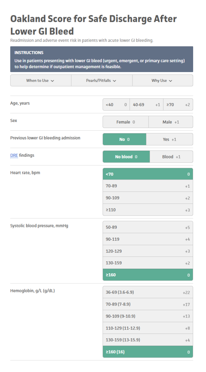
Screening tool for - Risk stratification - Oakland Score - Applicable for in-person evaluation
-
To identify low-risk patients with LGIB who are appropriate for early discharge and outpatient diagnostic evaluation. Risk scores should be used to supplement but not replace clinician judgment.
- The original score was created and validated using a cutoff of ≤8.
- Scores >8 identify high-risk patients who may need hospital admission.
-
Clinical judgment should always be utilized when determining disposition planning for patients with suspected lower GI bleed.
▶️ Initial Management
-
Triage
- Unstable / ongoing brisk bleeding → ED transfer, IV access, resuscitation, type & screen.
- Stable → risk stratify and proceed with diagnostic plan.
-
“Telemedicine Exclusion Criteria”
-
Clearly state conditions not appropriate for continued tele‑management:
- Ongoing brisk bleeding or recurrent episodes in 24 hours.
- Orthostasis, syncope, altered mental status.
- Known cirrhosis, active malignancy, recent colonoscopy with intervention.
- Inability to reliably communicate or follow instructions.
-
Clearly state conditions not appropriate for continued tele‑management:
-
Labs
- CBC, BMP , INR/PTT (if on anticoagulants), lactate if concern for hypoperfusion.
-
Imaging / Endoscopy
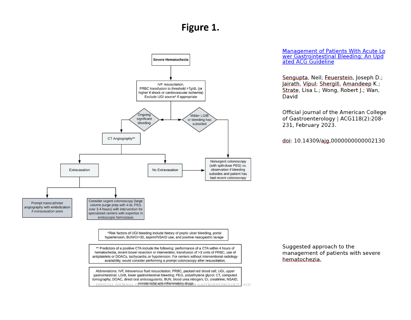
- Milder LGIB or bleeding has subsided -> Nonurgent colonoscopy (with split dose PEG) vs Observation if bleeding subsides and patient has had recent colonoscopy
-
Active, significant bleeding: CTA prioritized to localize source; institutional protocols improve process outcomes and reduce transfusions
- CTA as the initial diagnostic test in patients with ongoing hemodynamically significant hematochezia. However, CTA is of low yield in patients with minor LGIB or those in whom bleeding has clinically subsided. (Conditional recommendation, low-quality evidence)
- Stable major LGIB: admit for colonoscopy on next available list
- Recurrent bleeding after initial hemostasis: repeat colonoscopy with endoscopic therapy if indicated
-
Medical Management
- Hold offending agents when feasible (NSAIDs; manage antithrombotics case‑by‑case).
- Transfuse based on hemodynamics/clinical context (done if at ER)
-
After negative evaluation and stability, consider iron therapy if iron deficiency present
💆 Treatment (Etiology‑Directed)
- Diverticular bleed, angioectasia, post‑polypectomy, ischemic colitis, hemorrhoidal/anorectal sources—manage per endoscopic findings.
- Failure of endoscopy or inability to stabilize → radiology or surgical consultation
🔮 Follow Up & Surveillance
- No further bleeding, stable: outpatient follow‑up in 1–2 weeks; review pathology, iron studies, medication plan.
- Recurrent hematochezia: expedited GI follow‑up; consider repeat colonoscopy
-
Iron deficiency without source: monitor response to iron and reassess if recurrence occurs
🤝 Referral to Specialist (GI)
- Immediate GI consult: active bleeding, hemodynamic changes, need for endoscopic therapy.
- IR/Surgery: severe hematochezia unable to be stabilized or failed endoscopic localization/hemostasis
- Admission: all major LGIB for inpatient colonoscopy
</> Algorithm (Clinic → ED → Inpatient)
Hematochezia → Assess stability
- If unstable/brisk → ED → resuscitate + labs → CTA → targeted endoscopy/IR → surgery if needed.
- If stable → labs + rectal exam → risk stratify
- Major LGIB → Admit → colonoscopy next available.
- Minor/self‑limited → outpatient GI plan.
- After hemostasis → monitor → if rebleed → repeat colonoscopy → escalate to IR/surgery if refractory.
If you have any questions or would like more information, please reach out to the Delegating Physician.
Comments (0)