
📚Etiology
-
Viral (most common): Rhinovirus, influenza, parainfluenza; typically follows URI.
- Acute viral rhinosinusitis (AVRS) begins with viral inoculation via direct contact with the conjunctiva or nasal mucosa
- Viral rhinitis spreads to the paranasal sinuses
-
Bacterial (ABRS, minority): Streptococcus pneumoniae, Haemophilus influenzae, Moraxella catarrhalis.
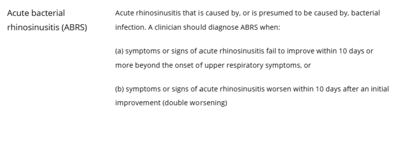
- Acute bacterial rhinosinusitis (ABRS) occurs when bacteria secondarily infect an inflamed sinus cavity.
- Noninfectious contributors: Allergic rhinitis, irritant exposure, anatomic obstruction.
Guidelines emphasize that most adult cases are viral and self-limited
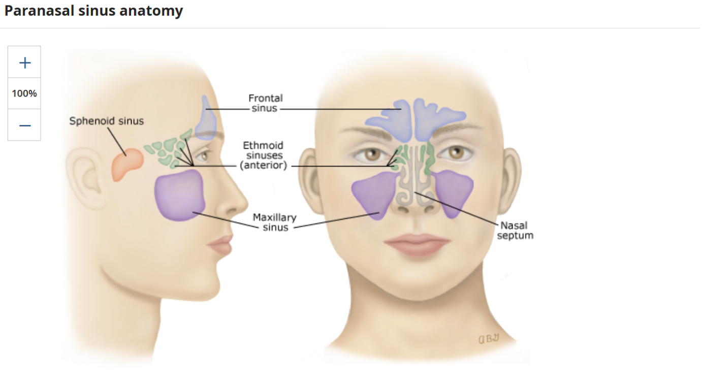
Classification by duration
- <4 weeks = acute
- 4-12 weeks = subacute
-
>12 week = chronic
- The healthcare provider cannot diagnose chronic sinusitis based on symptoms alone. The healthcare provider will also need to see nasal swelling, nasal drainage, or inflammation on exam.
- CT scan may confirm a diagnosis of chronic sinusitis or inflammation
Recurrence Classification
-
Recurrent = 4 or more annual episodes without persistent symptoms between episodes
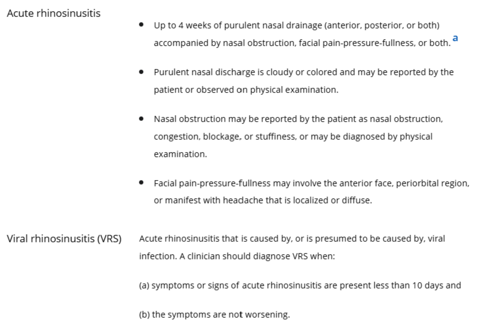
🦠 Pathogenesis
- URI → nasal mucosal inflammation → sinus ostial obstruction.
- Impaired mucociliary clearance → mucus retention.
- Secondary bacterial overgrowth may occur in a subset of patients after prolonged or worsening illness.
📹 Telemedicine HPI Intake
Focused symptom-based intake is sufficient for diagnosis.
- Duration and trajectory of symptoms
-
Key symptoms
- Nasal obstruction or purulent nasal discharge
- sinus infection, there is usually cloudy or colored nasal drainage.
- Facial pain/pressure (worse bending forward)
- Hyposmia/anosmia
- Systemic features: Fever, malaise
-
Pattern flags
- ≥10 days without improvement
- “Double worsening” after initial improvement
-
Severity markers
- Pain severity
- Fever ≥38.3°C (101°F)
-
Risk modifiers
- Immunocompromise
- Recent antibiotics
- Recurrent episodes (≥4/year)
Clinical diagnosis is history-based; physical exam findings are not required for
uncomplicated cases
CT scan is NOT needed to diagnose acute sinusitis**
🚩 Screening for Red Flags
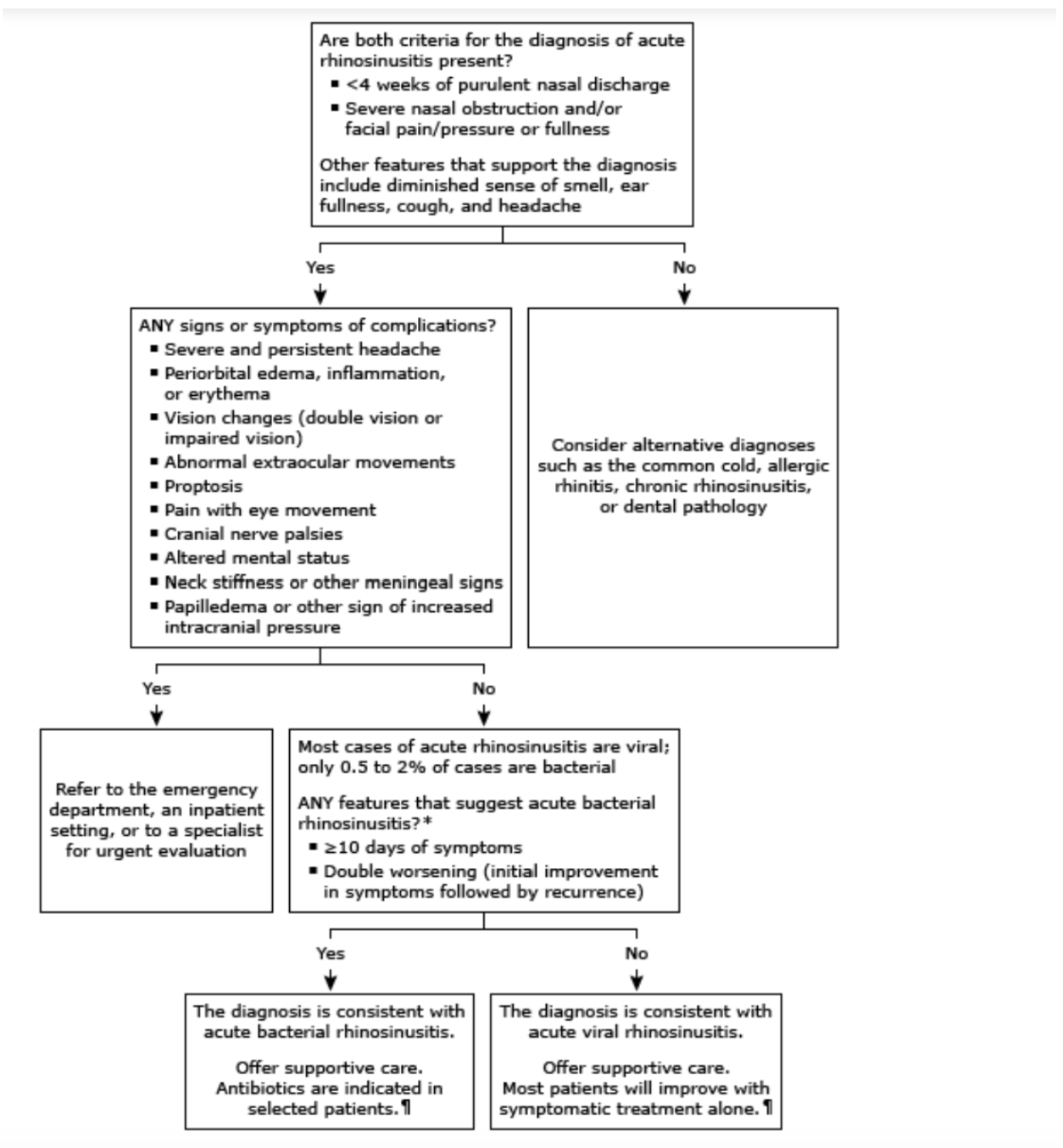
Immediate escalation if any of the following are reported:
- Periorbital edema, erythema, or vision changes
- Severe headache, altered mental status
- High fever with systemic toxicity
- Focal neurologic deficits
- Signs of orbital or intracranial complication
These findings warrant urgent in-person evaluation or ED referral
Possible Complications
- Orbital cellulitis
- Preseptal cellulitis
- Intracranial abscess
- Menigitis
🩻 Testing & Imaging
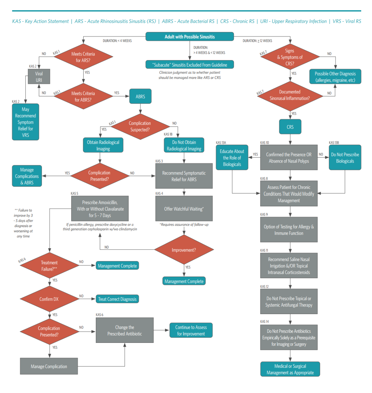
No routine labs or imaging for uncomplicated ARS diagnosed clinically.
CT or imaging is contraindicated unless complications or alternative diagnoses are suspected
Viral or bacterial testing is not indicated in routine adult ARS.
🎯 Diagnosis (Telemedicine Criteria)
-
Acute Viral Rhinosinusitis
- Symptoms <10 days and improving
- Acute Bacterial Rhinosinusitis (ABRS)
-
Diagnose when either is present:
- Symptoms ≥10 days without improvement
- Worsening symptoms after initial improvement (“double worsening”)
This distinction is a strong guideline recommendation
💆 Management & Treatment
Initial Symptomatic Management (All Patients)
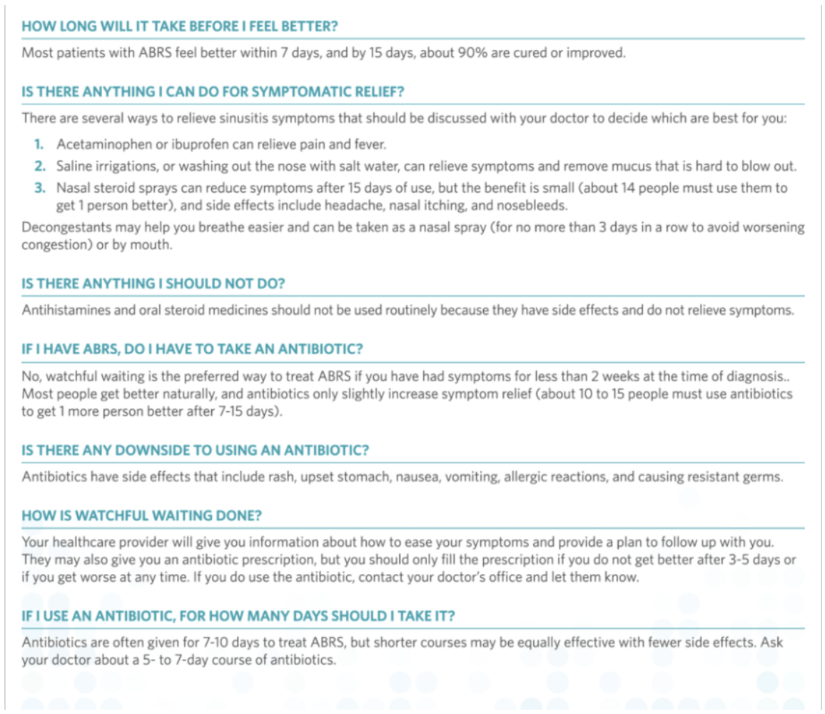
- Analgesics - acetaminophen, NSAIDs
- Intranasal corticosteroids
- Saline nasal irrigation
Symptomatic therapy is appropriate for viral ARS and ABRS
Antibiotic Strategy (ABRS Only)
- Option 1: Watchful waiting
- Mild symptoms, reliable follow-up
- Option 2: Antibiotics
- First-line: Amoxicillin ± clavulanate
- Duration: 5–10 days
- Reassess at 7 days
- If no improvement or worsening → confirm diagnosis, adjust therapy, or escalate care
Watchful waiting and short-course therapy are guideline-supported to reduce overtreatment.
🔮 Follow Up
- Asynchronous or synchronous check-in at 7 days
- Document symptom trajectory and adherence
-
Reinforce return precautions for complications
🤝 Referral to Specialist (ENT)
Refer when any of the following are present:
- Suspected orbital or intracranial complication
- Recurrent acute rhinosinusitis (≥4 episodes/year)
- Chronic rhinosinusitis symptoms >12 weeks
- Failure of appropriate medical therapy
- Concern for anatomic obstruction or nasal polyps
ENT evaluation may include endoscopy or CT imaging when indicated
If you have any questions or would like more information, please reach out to the Delegating Physician.
Comments (0)